


Livestock Diseases
Salmonella contamination in animal-derived-meals used in feed manufacturing
- Abstracts: English Portuguese
Abstract in English:
From three feed factories in the State of São Paulo, Brasil, 204 samples of animal-derived meals were collected, of which 158 were meat-meals, 25 were feather-meals, 16 were feather and viscera-meals, three were viscera-meals and two were bone-meals. All of them were-t:o be employed in feed mixtures. The samples were bacteriologically processed and from the total 204, as many as 77 were contaminated with salmonellae, from one to six serotypes being identified in individual samples. The proportion of infected samples were respectively: 39% of the meat-meals, 16% of the feather-meals, 63% of the feather and viscera-meals, 33% of the viscera-meals and 50% of the bone-meals. The most frequent represented serotypes were Salmonella cerro (19 samples) and S. havana (17 samples). Better results were obtained by the use of selenitenovobiocin broth than by employement of tetrathionate-novobiocin broth, in both cases after a pre-enrichment in Ringer 1/4.
Abstract in Portuguese:
Foram analisadas 204 amostras de farinhas de origem animal, empregadas na elaboração de rações em três fábricas situadas no Estado de São Paulo, correspondendo a 158 amostras de farinha de carne, 25 amostras de farinha de pena, 16 amostras de farinha de pena e víscera, três amostras de farinha de vísceras e duas amostras de farinha de osso. Das 204 amostras, 77 estavam contaminadas por Salmonella. Essas 77 amostras, individualmente albergavam salonelas de um a seis sorotipos. O percentual de positividade foi de 39%, 16%, 63%, 33% e 50%, respectivamente, para as farinhas: de carne, de pena, de pena e víscera, de víscera e osso. Os sorotipos mais freqüentes foram Salmonella cerro (19 cepas) e S. havana (17 cepas). Melhores resultados foram conseguidos a partir do caldo selenito-novobiocina, com relação ao caldo tetrationatonovobiocina, após pré-enriquecimento das amostras em solução de Ringer 1/4.
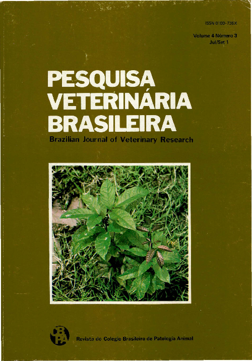
Experimental poisoning by Arrabidaea bilabiata (Bignoniaceae) in rabbits
- Abstracts: English Portuguese
Abstract in English:
Dried and powdered leaves of Arrabidaea bilabiata (Sprague) Sandw. (fam. Bignoniaceae), a plant toxic for cattle, were administered by stomach tube to 57 rabbits, in doses that ranged from 0.5 to 6.0 gtams per kilogram of body weight. Twenty-six of the rabbits died. A large variation in the dose that caused symptoms and death of the animals was observed, the smallest dose that killed rabbits being 1 g/kg, while the largest that did not was 6 g/kg. Three rabbits that received 0:5 g/kg did not die. Death occurred in two of the eight rabbits that were fed 1 g/kg, in eight of 16 receiving 2 g/kg, in four of 12 that received 4 g/kg, and in 10 of 16 that were administered 6 g/kg of body weight. In the present experiments, the first symptoms were noted froin 2h22' to 12h07' after ingestion of the plant. The course of the poisoning lasted from half a minute to 4 minutes, although in one case it lasted 17 minutes. The main symptonis were those of "sudden death": the rabbits, in general, made sudden violent, uncontroiled movements, struggle or jumped, while at other times only slow uncontrolled movements were observed, after which, the animals generally fell on their sides. Some animals just fell on their sides. Respiration became difficult and intermitente and the animals died. From the begfuning of syniptoms untíl death the rabbits screamed frequently. Af necropsy, no gross lesions were. found. Microscopic lesioris were present in the tiver, kidney and heart, and were essentially·degerierative and vascular in·nature. In the liver there was necrosis, vacuolization of the cytopla Sma and albuminous-granular degeneration of hepatocytes, congestion, dissociation of the liver cords, compressive atrophy of these, presence of eosinophilic sphaerules in the sinusoids and edema of Disse’s space. In the kidney hydropic-vacuolar degeneration of the epithelial cells of the distal convoluted tu bules and swelling of the epithelial cells of the convoluted tu bules at the cortico-medular junction was found. Heart lesions were characterized by interacellular edema of the heart fibres, dissociation of them and foci of increased eosinophilia of the heart muscle.
Abstract in Portuguese:
As folhas dessecadas e pulverizadas de Arrabidaea bilabiata (Sprague) Sandw. (fam. Bignoniaceae), planta tóxica para bovinos, foram administradas por via intragástrica em quantidades que variaram de 0,5 a 6 g/kg, a 57 coelhos. Desses, 26 morreram. Houve grande variação na dose que causava o aparecimento de sintomas e a morte dos animais. A dose menor que causou a morte dos coelhos foi 1 g/kg;·a maior que não matou coelhos foi 6 g/kg. Nenhum dos 3 coelhos que receberam 0,5 g/kg morreu. De 8 coelhos que receberam 1 g/kg, 2 morreram; de 16 coelhos que receberam 2 g/kg, 8 morreram; de 12 coelhos que receberam 4 g/kg, 4 morreram; e de 16 coelhos que receberam 6 g/kg, 10 morreram. O início dos sintomas variou de 2h22' a 12h07' após o início da administração da planta. A evolução da intoxicação variou de meio a 4 minutos; em um coelho foi de 1 7 minutos. Os sintomas de intoxicação foram os da síndrome de "morte súbita": repentinamente o coelho fazia movimentos desordenados, violentos, debatia-se ou pulava; outras vezes só fazia subitamente movimentos desordenados lentos e em seguida caía, em geral, em decúbito lateral. Outros simplesmente caíam; também em decúbito lateral. A respiração tornava-se difícil, espaçada e o animal morria. Desde o início do aparecimento dos sintomas até a morte, a maioria dos coelhos emitia gritos, com maior ou menor freqüência. Os achados de necropsia foram praticamente negativos. Os exames histopatológicos revelaram alterações no fígado, rim e coração, consistindo principalmente em alterações degenerativas e vasculares. No fígado foram observadas necrose, vacuolização do citoplasma e degeneração albuminosa-granular dos hepatócitos, congestão, dissociação dos cordões hepáticos, atrofia compressiva desses, presença de esférulas eosinofílicas nos sinusóides e edema dos espaços de Disse; no rim, degeneração hidrópico-vacuolar das células epiteliais dos túmulos contorna dos distais e·tumefação das células epiteliais dos túbulos contornados na junção Cortico-medular: no coração edema intracelular das fibras cardíacas, afastamento entre estas e presença de focos de eosinofilia aumentada do músculo cardíaco.
Vaccination of chickens against chronic respiratory disease with an oil-emulsion Mycoplasma gallisepticum bacterin
- Abstracts: English Portuguese
Abstract in English:
Subcutimeous inoculation of 30-day old SPF chickens with a·commercial oilaemulsfon Mycoplasma gallisepticum bacterin (MG-Bac, lot 23009, Salsbury Lab., Inc., Charles City,·Iowa 50616, USA) protected them against chronic respiratory disease. The·birds were challenged 30 days after vaccination with the R strain of M. gallisepticum injected into the left thoracic air·sac. The vaccinated birds presented, before and after the challenge, geometric mean titers higher than the nonvaccinated ones. Air sacculitis lesions bccurred with lower frequency and intensity among the vaccinated birds than among the nonvaccinated, showing a positive relationship between the protection and the serological response.
Abstract in Portuguese:
A inoculação subcutânea de galinhas "SPF" de 30 dias de idade com uma vacina oleosa comercial de Mycoplasma gallisepticum (MG-Bac, partida 23009, Salsbury Lab., Inc., Charles City, Iowa 50616, EUA) protegeu-as contra a doença respiratória crônica. As aves foram desafiadas 30 dias após a vacinação pela injeção no saco aéreo torácico esquerdo com a amostra R de M gallisepticum. As aves vacinadas apresentaram, antes e após o desafio, médias geométricas dos títulos de inibição da hemaglutinação maiores do que as aves não vacinadas. As lesões de aerossaculite ocorreram com frequência e intensidade menores entre as aves vacinadas, relacionando-se positivamente proteção obtida com a resposta sorológica observada.
Equine leukoencephalomalacia in horses in Rio Grande do Sul, Brazil
- Abstracts: English Portuguese
Abstract in English:
An outbreak of equine leukoencephalomalacia, associated with moldy corn poisoning occuring during the months of Juné-July in Rio Grande do Sul, is described. Ten horses from 4 different farms in Lavras do Sul county were affected. Clinical signs irtcluded anorexia, circling, head pressing, blindness, ataxia, recumbency and death. All affected horses died within a period of 13-22 hours after the onset of clinical signs. Post-mortem examination was performed in 3 horses. Lesions were restricted to the central nervous system. The most striking gross lesions were focal areas of softening and cavitation of the cerebral white matter. These areas were surroundeci by multiple minute hemorrhagic foci. Histopathological examination revealed these areas of cavitation to consist of hollow spaces surrounded by malacic, hemorrhagic and edematous neuropile. Other microscopic brain lesions included generalized hypertrophic and degenerative changes of the vascular endothelia, perivascular hemorrhages and edema; presence of perivascular eosinophilic globules; edema of the white matter; sludging, margination and pavimentation of leukocytes and mild perivascular cuffings consisting mainly of polimorphonuclear granulocytes, neutrnphils and eosinophils. Fusarium moni/iforme was isolated from the corn used to feed the horses: It is suggested that a primary vascular change is the early event in the pathogenesis of the overt malacic foci.
Abstract in Portuguese:
É descrito um surto de ·leucoencefalomalácia associada a ingestão de milho mofado que ocorreu em equinos durante os meses de junho-julho de 1983 no Rio Grande do Sul. Dez cavalos oriundos de 4 diferentes estabelecimentos no município de Lavras do Sul foram afetados. Os sinais clínicos consistiam em anorexia, andar em círculos, pressão da cabeça contra objetos, cegueira, ataxia, decúbito e morte. Todos os eqüinos afetados morreram de 13 a 22 horas após o início dos sintomas. Foram realizadas necropsias em 3 animais. As lesões encontradas restringiam-se ao sistema nervoso central. A lesão macroscópica mais evidente era representada por áreas focais de amolecimento e cavitação da substância branca do cérebro. Essas áreas eram cercadas por vários pequeninos focos hemorrágicos. Ao exame histopatológico, essas áreas consistiam em cavidades vazias cercadas por tecido nervoso necrótico-hemorrágico e edematoso. Outras alterações microscópicas observadas no cérebro foram alterações hipertróficas e degenerativas generalizadas do endotélio vascular, hemorragias e edema perivasculares, presença de glóbulos eosinofílicos no espaço de Virchow-Robin, edema da substância branca, marginação e pavimentação leucocitária e discretos manguitos perivasculares constituídos principalmente de neutrófilos e eosinófilos. O fungo Fusarium moniliforme foi isolado do milho obtido da alimentação dos cavalos. Sugere-se que uma alteração vascular primária seja o evento inicial na patogenia das áreas de malácia.
Virulence factors present in cultures of Escherichia coli isolated from pigs in the region of Concórdia, Santa Catarina, Brazil
- Abstracts: English Portuguese
Abstract in English:
Four hundred and seventy-seven cultures of Escherichia coli isolated in the region of Concórdia, Santa Catarina, Brazil, were examined for the presence of ''virulence factors", such as: thermolabile (LT) enterotoxin; thermostable (STa and STb) enterotoxins and the colonization factors K88, K99 and 987P. Paired samples of sera from some sick piglets· were also collected for the search of anti-LT antibodies. Eighty-six (18,02%) E. coli cultures produced ·STa enterotoxin whereas LT enterotoxin was detected in 8 (1,67%) only. Among 391 STa- cultures 49 (12,53%) were STb+. Among 28 K88+ cultures, 1 produced LT; 9 STa and the remaining were non-enterotoxigenic. None of the L T+ cultures codified for K99 antigen, which, on the other hand, was found in 5 STa+ and in 17 non-entérotoxigenic cultures. 987P antigen was found in 3 non-enterotoxigenic cultures. Four out of 10 samples of the examined paired sera showed serological conversion in relation to titres found in ·the passive immunehemolysis (PIH) test. The results of serogrouping revealed that all LT+ cultures belonged to serogroup 01.49. Among 86 STa+ cultures, 1 was grouped as serogroup 09, 2 as 010, 4 as 035; 2 as 064; 3 as 0108; 2 as 0138; 1 as 0149 and 1 as 0157. Seventy STa+ cultures were not classified.· Among 49 STb+ cultures 1 belonged to serogroup 09; 4 to 010; 1 to 035; 1 to 0139; 2 to 0149; 1 to 0157 and 39 cultures could not be serogrouped. These results suggest that in porcine colibacillosis of the region of Concórdia, SC, Brasil the thermostable enterotoxins (STa and STb) may play an important role. It could be possible that among these cultures other colonization factors, different from K88, K99 and 987P may occur. Also, most cultures examined could not be grouped among the serogroups generally accepted as enteropathogenic for swine.
Abstract in Portuguese:
Quatrocentos e setenta e sete amostras de Escherichia coli, isoladas na região de Concórdia, SC, Brazil, foram examinadas quanto a presença de fatores de virulência a saber: enterotoxína termolábil (LT), enterotoxinas termoestáveis (STa e STb) e os fatores de colonização K88, K99 e 987P. Amostras pareadas de soro de alguns leitões doentes foram também coletadas para a pesquisa de anticorpos anti-LT. Oitenta e seis (18,02%) amostras de E. coli produziram a enterotoxina STa enquanto a enterotoxina LT-foi detectada em apenas 8 (1.67%). Entre as-381 amostras STa-, 49 (12.53%) foram STb+. Entre 28 amostras K88+, uma produziu LT, 9 STa e as restantes eram não enterotoxigenicas. Nenhuma das amostras ·LT+ codificou para o antígeno K99, porém, este foi encontrado em 5 amostras STa+ e em 17 não entérotoxigênicas. Quatro entre os 10 soros examinados apresentaram conversão sorológica nos níveis de anticorpos anti-LT quando os soros foram examinados pelo teste da imunohemólise passiva (PIH). Os resultados dos exames sorológicos revelaram que todas as amostras LT+ pertenciam ao sorogrupo 0149. Entre 86 amostras STa+, 1 foi classificada como pertencendo ao sorogrupo 09, 2 ao 010; 4 ao 035; 2 ao 064; 3 ao 0108; 2 ao 0138; 1 ao 0149; 1 ao 0157 e 70 amostras não foram Classificadas. Entre as 49 amostras STb+ 1 pertencia ao sorogrupo 09; 4 ao 010; 1 ao 035; 1 ao 0139; 2 ao 0149; 1 ao 0157 e 39 não foram classificadas. Estes resultados sugerem que na colibacilose suína da região· de Concórdia as enterotoxinas termoestáveis (STa e STb) possam desempenhar um importante papel, existindo a possibilidade de que nelas possam ocorrer outros fatores de colonização que não K88, K99 e 987P. As amostras estudadas também, em sua maioria, não se enquadraram entre· aqueles sorogrupos geralmente aceitos como enteropatogênicos para suínos.
