


Livestock Diseases
Surgical and non-surgical embryo transfer in cattle
- Abstracts: English Portuguese
Abstract in English:
From October through December, 1980, a study of embryo transfer in cattle was carried out in the State of Rio Grande do Sul, Brazil. Twenty-five cows were used as donors. The breeds involved were Hereford (10), Aberdeen Angus (4), Normand (6) and Charolais (5). Forty cows Hereford Aberdeen Angus, Normand, Charolais and Holstein crosses were used as recipients. Oestrus synchronization and superovulation were obtained using prostaglandins alpha analogue and PMSG. Fourteen (56%) of the donors responded well to the superovulation treatment with PMSG, producing an average of 6.2 corpora lutea per animal. Thirty-eight embryos were collected by the non-surgical method, 29 of these being normal in development and it morphology normal. After 21 transfers using the surgical method 15 (71%) recipients became pregnant. Two out of 6 (33%) recipients treated by the non-surgical method sustained the pregnancy. It is concluded that the surgical method for bovine embryo transferis still the most efficient. However, the need for rapid improvement is the non-surgical method is stressed, due to its simplicity and easy manipulation in the field.
Abstract in Portuguese:
Durante outubro e dezembro de 1980 foi desenvolvido um estudo na área de transferência de embriões em bovinos no Rio Grande do Sul. Como doadoras foram utilizadas 25 fêmeas das raças Hereford (10), Aberdeen-Angus (4), Normanda (6) e Charolesa (5). Para receptoras foram selecionadas vacas cruza Holandesa e das raças supracitadas. Obteve-se a sincronização do ciclo estral e superovulação através da aplicação de um análogo de prostaglandina 7 F2 alfa e de PMSG8. Das 25 vacas superovuladas 14 (56%) responderam ao tratamento com PMSG com uma média de 6,2 C.1./animal. Foram coleta- dos 38 embriões pelo método não cirúrgico, sendo 29 (76% com morfologia e desenvolvimento normais. Após 21 transferências pelo método cirúrgico, 15 (71%) receptoras resultaram gestantes; após 6 pelo não cirúrgico 2 (33%) receptoras mantiveram a gestação. Conclui-se ainda que o método de transferência cirúrgica é o mais eficiente e que há a necessidade de acelerar-se o aperfeiçoamento do método de transferência não cirúrgico em função da simplicidade e rapidez da manipulação para trabalhos a nível de campo.
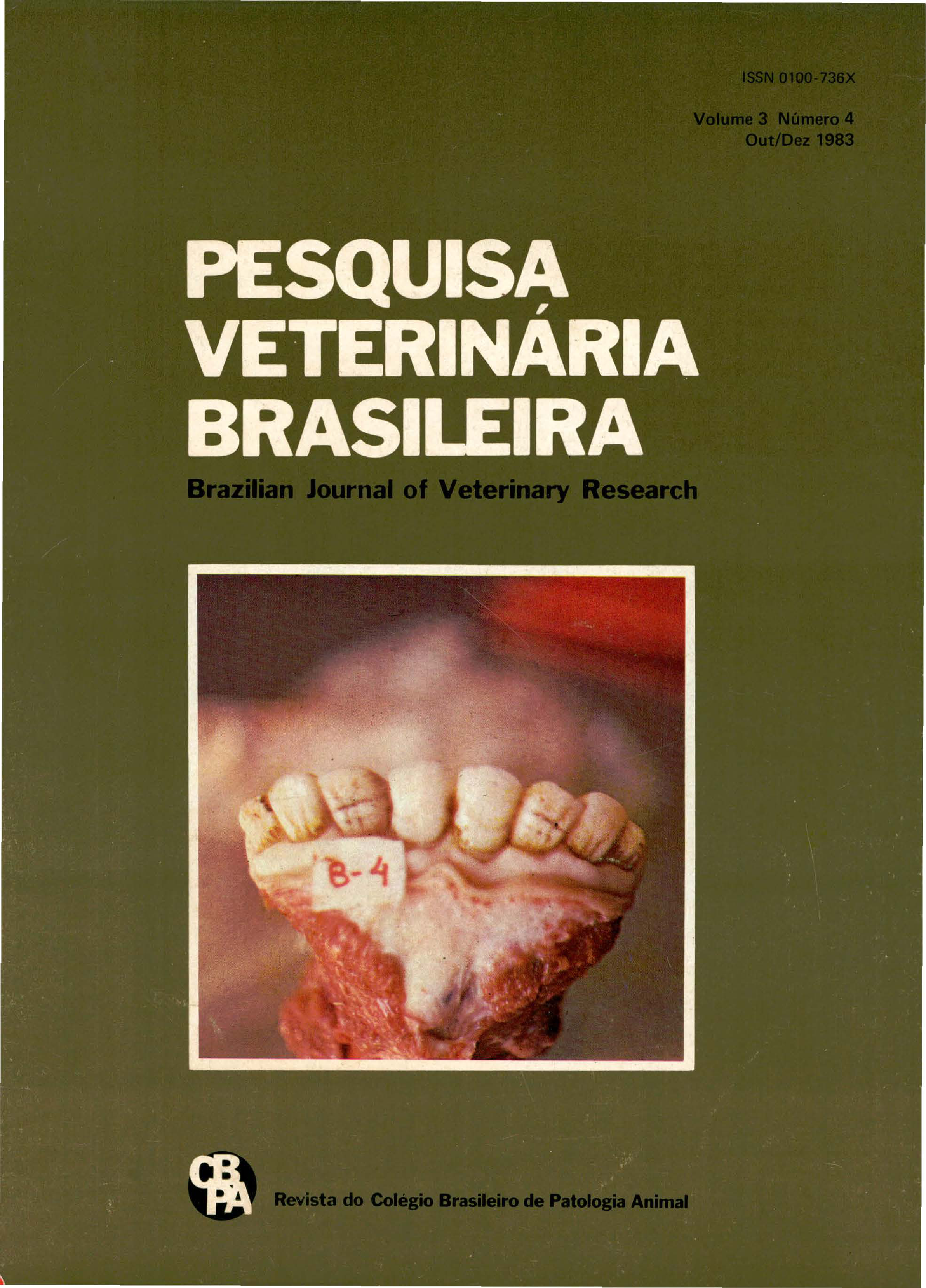
Industrial pollution as a cause of fluoride intoxication in cattle in the municipality of Rio Grande, southern Brazil
- Abstracts: English Portuguese
Abstract in English:
Fluoride intoxication in cattle occurred as a consequence of atmospheric pollution caused by the industrial effluents of 4 phosphate processing factories located in the city of Rio Grande, state of Rio Grande do Sul. The lesions of the incisor teeth were characterized by a chalky white or brown discoloration, hypoplasia of the enamel, increased attrition and gingival hyperplasia. Such lesions were studied in animals from 19 farms loca te d in a range of 4 .5 to 17 .5 km from the factories. The severity of the lesions was related to the distance between the farms and the factories as a linear function [Y = 2.13 + (- 0.12. x); r2 = 0.77 (P < 0.001)]. Molar and premolar teeth showed excessive and uneven attrition. In a farm located 6 km from the factories, 2 cows showed lameness and 1 had hyperostosis of the metatarsal bones. Histological lesions of the permanent incisor teeth were hyperplasia of the cement and disturbed incremental lines and hypocalcification of the dentine. Bone lesions were characterized by osteoporosis with degeneration and atrophy of osteoblasts. Numerous cementing lines and retention of the chondroid core were observed, indicating arrested osteocytic osteolysis and chondrolysis. In compact bone, osteons were irregular in shape, size and distribution with enlarged Haversian canals and irregular distribution of osteocytes; insterstitial lamellae were increased and irregular. It is considered that the lesions of osteofluorosis are due to an imbalance between bone apposition and resorption; osteoporosis would be a result of the alterations observed in osteoblastos which were apparently more severely affected than osteocytes. Fluoride leveis, determined in the epifise-metafise of the humerus of 3 animals and in the mandibles of 7 belonging to two properties situated between 5 and 6 km from the factories, varied between 1,400 ppm and 5,750 ppm of ash. This paper points out the potential risks of fluoride contamination by the human population of the city of Rio Grande, where the fosfate processing factories are situated.
Abstract in Portuguese:
Descreve-se intoxicação por flúor, em bovinos, como conseqüência da poluição atmosférica produzida por fábricas de adubo que processam rocha fosfática, localizadas na cidade de Rio Grande, Rio Grande do Sul. As lesões dos· incisivos caracterizaram-se por manchas esbranquiçadas com aspecto de giz, pigmentação marrom, hipoplasia do esmalte, desgaste dentário exagerado e hiperplasia da gengiva. Tais lesões foram estudadas em 19 estabelecimentos localizados entre 4,5 e 17 ,5 km de distância das fábricas de adubo, determinando-se uma função linear do grau das lesões com relação à distância [Y = 2,13 + (-0,12 . x); r2 = 0,77 (P < 0.001)]. Os molares e pré-molares apresentaram desgaste excessivo e irregular. Em um estabelecimento, localizado a 6 km das fábricas, foram observadas lesões de hiperostose em um animal, e claudicação em dois. Histologicamente as lesões dentárias foram caracterizadas por hiperplasia do cemento assim como distúrbios das linhas incrementais e hipocalcificação da dentina. As lesões ósseas consistiram em osteoporose com atrofia e degeneração dos osteoblastos; foram observadas também retenção da corda condróide e presença de linhas cimentantes, evidenciando uma inibição da condrólise e osteólise osteocítica. No osso compacto, os sistemas de Havers apresentavam-se irregulares em seu tamanho, forma e distribuição, os canais de Havers estavam aumentados de diâmetro e os osteócitos irregularmente distribuídos dentro do sistema; as lamelas intersticiais estavam aumentadas e irregulares. Considera-se que as alterações ósseas foram produzidas por um desequilibrio entre a aposição e a reabsorção óssea. A osteoporose teria ocorrido como consequência das lesões dos osteoblastos, os quais foram aparentemente mais afetados que os osteócitos. Os níveis de flúor, determinados em 7 mandíbulas e 3 úmeros de animais pertencentes a dois estabelecimentos situados entre 5 e 6 km das fábricas, variaram entre 1.400 e 5 .750 ppm. Alerta-se para os riscos de saúde, pela contaminação por flúor, a que estão expostos os habitantes da cidade de Rio Grande, onde se localizam as fábricas processadoras de rocha fosfática.
Experimental poisoning of cattle by Crotalaria anagyroides (Leg. Papilionoideae)
- Abstracts: English Portuguese
Abstract in English:
Symptoms of toxicity were observed in calves fed fresh aerial parts of the plant Crotalaria anagyroides collected in the State of Rio de Janeiro. Plant material was given during periods ranging from 1 to 12 days, the amount consumed by each animal varying from day to day. The plant was not well eaten and the original plan of giving it as the only food could not be adhered to. ln most experiments plant material was mixed with small amounts of chopped giass, concentrate ration and molasses; at times it was necessary to place the plant in the mouth of the animals. The amounts eaten varied from 6700 g (55.8 g/kg) eaten by one animal on one day, to 55400 g (294.8 g/kg) eaten by another animal over 12 days. Two animals received only the leaves and thin stems, two others only the flowers and green and ripe pods, and the last two, all aerial parts (leaves, thin stems, flowers and green and ripe pods). All calves suffered the toxic effects of the plant. Five of the six calves had clinical symptoms of poisoning, while the sixth showed only histopathologic lesions. Symptoms of poisoning were first seen between 38 days and 9 months and 8 days after the first day of ingestion of the plant, until which time the animals were apparently healthy and in good nutritional state. Symptoms consisted mainly of a lack of appetite and weight loss, over weaks or months (3/5) and/or nervous symptoms as excitation (2/5) or depression (1/5) which lasted for days. Two animals also had abdominal contractions of short duration. Two animals died, 11 days and 35 days after the onset of symptoms, one was euthanazied in extremis, after 9 days of disease, and another was euthanazied in bad nutritional state, after 4 months of disease. Only one animal recovered clinically, having shown symptoms for six months and then regaining weight. It was euthanazied together with the sixth bovine which had never shown symptoms of poisoning. The most important post-mortem finding was cirrhosis: the liver was hard, pale and had a lobular, granular or nodular aspect. The most important histopathological changes were the presence of collagen, proliferation of fibroblasts and of the epithelial cells of the bile ducts, megalocytosis and the presence of nodules of regeneration in the liver; in two animals spongy degeneration in the central nervous system. Attention is drawn to the fact that no case histories have been obtained or observations made that incriminate Crotalaria anagyroides as the cause of spontaneous disease in cattle, and it is believed that this would hardly happen because of the bad palatability of the plant.
Abstract in Portuguese:
Em experimentos em que as partes aéreas de Crotalaria anagyroides frescas, recém-colhidas na área do Km 47, Seropédica, município de Itaguaí, Estado do Rio de Janeiro, foram administradas por via oral a seis bovinos jovens desmamados, a planta demonstrou ser tóxica para essa espécie animal. As folhas e talos finos, as inflorescências e vagens ou todas as partes juntas foram dadas aos animais em quantidades que variaram de 6700 g (55,8 g/kg) em um dia até 55400 g (294,8 g/kg) em 12 dias. Todas as partes da planta se revelaram tóxicas. Cinco dos seis bezerros mostraram sintomas de intoxicação; o sexto mostrou a ação tóxica da planta somente através de lesões histopatológicas. Os sintomas apareceram de 38 dias a 9 meses e 8 dias após a ingestão da planta, mantendo-se os animais até então aparentemente sadios e em bom estado de nutrição; consistiram principalmente em anorexia e emagrecimento durante semanas a meses (3/5) e/ou sintomas nervosos sob forma de excitação (2/5) ou depressão (1/5) durante período mais curto (dias). Dois animais tiveram também contrações abdominais. Dois animais morreram após 11 e 35 dias de doentes, um foi sacrificado in extremis após 9 dias doente, e outro foi sacrificado em estado de nutrição precário após 4 meses doente. Só um animal se recuperou clinicamente e engordou, após 6 meses de sintomatologia; esse bovino foi sacrificado junto com o que nunca tinha mostrado sintomas de intoxicação. O achado de necropsia mais importante foi cirrose hepática: o fígado era mais duro, tinha coloração alaranjada clara e aspecto lobular, granular ou até nodular. Os achados histopatológicos mais importantes foram presença de tecido colágeno, proliferação de fibroblastos e das células epiteliais das vias biliares, megalocitose e a presença de nódulos de regeneração no fígado; em dois animais foi constatada degeneração esponjosa no sistema nervoso central. Uma vez que não foram obtidos históricos nem feitas observações que Crotalaria anagyroides tenha sido a causa de doença espontânea em bovinos, é externada a opinião que em virtude da má palatabilidade, que durante os experimentos a planta demonstrou possuir, isto dificilmente poderá acontecer.
Prevalence of enzootic bovine leukosis virus in dahy cattle in Parana
- Abstracts: English Portuguese
Abstract in English:
A representative sample of blood from dairy cattle in the Sta te of Parana was tested for the presence of antibodies against the virus of enzootic bovine leukosis (EBL) and it was found that the general prevalence of the virus was 20.7%, in 40.8% of the surveyed farms. The higher prevalences of the virus were found in the areas of the State where the economic significance of dairy production is greater and this prevalence was also higher in the older animals and in those of the Holstein breeds.
Abstract in Portuguese:
Uma amostragem representativa de sangue do rebanho bovino produtor de leite do Estado do Paraná foi submetida à prova específica para verificação de anticorpos contra o vírus da leucose enzoótica bovina (LEB), tendo sido encontrado um índice geral de prevalência de 20,7% em 40,8% das propriedades pesquisadas. As maiores prevalências do vírus foram encontradas nas áreas do Estado onde a importância econômica da bovinocultura de leite é maior e esta prevalência foi maior nos animais mais velhos e naqueles das raças Holandesas.
Pathogenicity of a strain of Marek's disease herpes virus
- Abstracts: English Portuguese
Abstract in English:
The objective of the present work was to study the pathogenicity of a Brazilian strain of Marek's disease herpes virus (MDV). Chicks experimentally infected at one day of age developed antibody which was detected by the agar gel precipitation test four weeks later. Contactexposed chickens developed antibody after eight weeks. Antibody persisted through the 12th week, when the last assay was performed. Uninoculated control chickens reared in isolation remained free of antibody during the sarne period. Viral antigens were detected in impression smears of spleen, thymus and bursa of Fabricius of inoculated chickens and in those of spleen and bursa of Fabricius of contact-exposed chickens by using a direct immunofluorescent technique. No fluorescence was observed in impression smears of brain, liver, gonads or kidney of all chickens tested. Impression smears of all tissues from uninoculated control chickens were negative. Precipitating antibody against the herpes virus of turkeys was demonstrated in the blood, and tissue extracts of kidney, feather tips, spleen, thymus, bursa of Fabricius and gonads of inoculated and contact-exposed chickens. No antibody was found in similar tissue extracts from uninoculated control chickens. Lymphoid infiltration corresponding to types A, B and C and their combination were observed in the vagus nerves and in the nerves of the brachial and sciatic plexus of chickens experimentally inoculated with MDV. Lymphoid infiltrations in the skin, kidney and gonads, as well as lymphoid depopulation with cystic degeneration of the thymus and bursa of Fabricius, were observed in the inoculated chickens. Similar lesions were observed in the kidney, skin, and nerves of contact-exposed chickens. No microscopic lesions were present in the uninoculated control chickens. It is concluded that the strain of MDV studied induces clinical symptoms, macroscopic and microscopic lesions characteristic of the neural form of Marek's disease, stimulates the development of antibody in the serum, spreads horizontally, and gives rise to viral antigens and antibody detectable in tissue extracts.
Abstract in Portuguese:
O objetivo do presente trabalho foi estudar a patogenicidade de uma cepa brasileira do vírus herpes da doença de Marek (VDM). Aves experimentalmente infectadas com um dia de idade, desenvolveram anticorpos os quais foram detectados pelo teste de precipitação em gel de ágar, após quatro semanas. Aves expostas por contato desenvolveram anticorpos após oito semanas. Tais anticorpos persistiram até a 12ª semana, quando o experimento foi encerrado. Aves testemunhas (não inoculadas) mantidas em isolamento, permaneceram livres de anticorpos durante o mesmo período. Antígenos virais foram detectados em impressões de baço, timo e bursa de Fabricius de aves inoculadas e em impressões de baço e bursa de Fabricius de aves expostas por contato, através da prova de imunofluorescência direta. Fluorescência não foi observada em impressões de cérebro, fígado, gônadas e rins de todas as aves testadas. Todas as impressões de tecidos de aves testemunhas foram negativas. Anticorpos precipitantes contra o vírus herpes de perus (HVT) foram demonstrados no sangue e nos extratos de tecidos renal, pontas de penas, baço, timo, bursa de Fabricius e gônadas de aves inoculadas ou expostas por contato. Anticorpos não foram encontrados nos extrados dos mesmos tecidos, de aves testemunhas. Infiltrações linfocitárias correspondentes aos tipos A, B e C e suas combinações, foram observadas nos nervos vagos e nos plexos braquial e ciático de aves inoculadas experimentalmente com o VDM. Infiltrações linfocitárias na pele, rins e gônadas, assim como, despovoamento linfóide com degeneração cística do timo e da bursa de Fabricius, foram encontradas nas aves inoculadas. Lesões semelhantes foram também observadas nos rins, pele e nervos das aves expostas por contato. Lesões microscópicas não foram visualizadas nas aves testemunhas. Conclui-se que a cepa do VDM estudada, induz sintomas clínicos, lesões macro e microscópicas características da forma neural da doença de Marek, estimula o desenvolvimento de anticorpos no soro, dissemina-se horizontalmente e dá origem a antígenos virais e anticorpos detectáveis nos extratos de tecidos.

