


Livestock Diseases
Eradication of Aujeszky's disease virus from reproductive swine herds using the test-and-removal method of antibody-positive pigs
- Abstracts: English Portuguese
Abstract in English:
Five reproductive herds and an Artificial Insemination Center with swine that had precipitating and/or neutralizing antibody for Aujeszky's disease virus (ADV) were identified in the State of Santa Catarina. ADV was eradicated on the basis of repeated testing followed by identification and removal of swine with antibody to ADV. The rates of infection detected in these herds were, respectively, 17%, 1.5%, 21.1%, 2.1%, 5.2% and 0.4%. The identification of antibody positive swine was initially made utilizing the plate immunodiffusion (ID) test and later, using the micro serumneutralization test associated or not to the ID test. In three herds, ADV was eradicated after the removal of antibody-positive swine identified in the first testing, indicating lack of lateral spread of the ADV strains involved. In two herds, ADV was eradicated only after the removal of positive swine identified in the second testing, indicating little lateral spread of the viruses involved. In one herd, ADV was only eradicated after the removal of antibody-positive swine identified in the third testing. The intervals between consecutive testings, varied between four and 28 weeks. It is concluded that ADV can be eradicated from infected herds through repeated testing and the immediate removal of swine with antibody for ADV.
Abstract in Portuguese:
No Estado de Santa Catarina, foram identificados cinco plantéis de reprodutores e uma Central de Inseminação Artificial que possuíam suínos com anticorpos precipitantes e/ou neutralizantes para o vírus da doença de Aujeszky (VDA). A erradicação baseou-se na testagem repetida, identificação e remoção de suínos com anticorpos para o VDA. As taxas de infecção detectadas nos seis plantéis foram, respectivamente de: 17; 1,5; 21,1; 2,1; 5,2 e 0,4%. A identificação dos suínos com anticorpos foi realizada, inicialmente, utilizando-se o teste de imunodifusão em placa e, posteriormente, através do teste de soroneutralização em microplacas, associado ou não ao teste de imunodifusão. Após a primeira testagem, foi possível erradicar o VDA em três destes plantéis, através da identificação e remoção dos suínos com anticorpos, indicando a não ocorrência da disseminação lateral dos VDA envolvidos. Em dois plantéis, o VDA foi erradicado somente após a remoção de suínos positivos, identificados na segunda testagem, indicando escassa disseminação lateral dos vírus envolvidos. Em um plantel, o VDA somente foi erradicado após a remoção dos suínos com anticorpos, identificados na terceira testagem. Os intervalos entre testagens consecutivas, variaram entre quatro e 28 semanas. Concluiu-se que o VDA pode ser erradicado de plantéis infectados através de testagem repetida e a imediata eliminação de suínos portadores de anticorpos para o VDA.
Experimental poisoning by Vernonia mollissima (Compositae) in rabbits
- Abstracts: English Portuguese
Abstract in English:
The dried and powdered sprouts of Vernonia mollissima Don (fam. Compositae), a plant toxic for cattle, sheep and goats, were administered by stomach tube to 24 rabbits nine to 15 months after collection. The plant was shown to be toxic for this species also, but with much variation in the lethal dose: six grams per kilogram of body weight caused the death of three out of four animals; 4 g/kg-two of four; 2 g/kg-three of four; 1 g/kg-one of four; 0.5 g/kg-one of four; whereas with 0.25 g/kg all four rabbits survived. In all cases first symptoms were noted between one and eight days after administration of the plant. The course of the poisoning lasted from two to seven days in the ten lethal cases and from eight to 14 days in the three rabbits that showed moderate to severe symptoms but went on to recover. The main symptoms were anorexia with fewer smaller dark feces. These animals also drank less and urinated less. The main post-mortem findings were seen in the liver, kidneys and digestive tract. The color of the liver was lighter than normal and its lobulation could be distinguished; the kidneys were much lighter in color both inside and out; and congestion and hemorrhages were seen in the digestive tract, mainly in the stomach mucosa. Microscopic examination revealed a severe toxic nephrosis as the principal lesion in all 10 rabbits that died: a majority of the epithelial cells of the uriniferous tubules in the cortex suffered coagulation necrosis, with distrophic calcification in five cases. Severe lesions were also observed in the livers of all 10 rabbits: severe steatosis associated with an albuminous-granular-vesicular degeneration of all or the greater part of the liver parenchyma; the steatosis affected most heavily the liver cells on the periphery of the lobule and diminishing in intensity towards the center of the lobule where albuminous-granularvesicular degeneration predominated. These experiments show that the rabbit can be used as a small experimental animal in the continuation of studies of V. mollissima in regard to toxic principais and effects, but the great individual variation in sensibility to the toxic action of the plant among rabbits must always be kept in mind.
Abstract in Portuguese:
A brotação dessecada e pulverizada de Vernonia mollissima Don, planta tóxica a bovinos, ovinos e caprinos, administrada a 24 coelhos por via intragástrica, entre 9 e 15 meses após sua colheita, demonstrou possuir toxidez também para essa espécie animal; porém a dose letal variou muito: 6 g/kg causaram a morte de três de quatro coelhos, 4 g/kg, de dois de quatro coelhos, 2 g/kg, de três de quatro coelhos, 1 g/kg, de um de quatro coelhos, 0,5 g/kg, também de um de quatro coelhos; com 0, 25 g/kg os quatro coelhos usados sobreviveram. Os primeiros sintomas apareceram entre 1 e 8 dias após a administração da planta (casos letais e não letais). A evolução da intoxicação, nos 10 casos letais, foi de 2 a 7 dias, e nos 3 coelhos que adoeceram mas se recuperaram, de 8 a 14 dias. Os sintomas foram principalmente anorexia e diminuição das fezes (acentuada), da sede e da urinação. Os achados de necropsia localizaram-se principalmente no fígado, nos rins e no aparelho digestivo. O fígado era mais claro e percebia-se sua lobulação; os rins eram bem mais claros, tanto na superfície como ao corte, e no aparelho digestivo se observaram, principalmente na mucosa do estômago, congestão e hemorragias. Os exames histopatológicos revelaram em todos 10 coelhos que morreram, como alteração mais importante, grave nefrose tóxica, sob forma de necrose por coagulação das células epiteliais da maior parte dos túbulos uriníferos da cortical, em 5 casos com calcificação distrófica; no fígado também foram constatadas alterações graves, em todos 10 coelhos, consistindo em acentuada esteatose associada a uma degeneração albuminosa granular vesicular afetando todo ou a maior parte do parênquima hepático; a esteatose afetava em grau acentuado as células hepáticas na periferia do lóbulo, diminuindo em intensidade em direção ao centro do lóbulo, onde predominava a degeneração albuminosa granular vesicular. Concluiu-se que o coelho pode ser usado como animal experimental de pequeno porte na continuação dos estudos sobre a ação tóxica de V. mollissima, bem como nos trabalhos de identificação de seus princípios ativos, mas é preciso sempre levar em consideração a grande variaçao na sensibilidade dos coelhos à ação tóxica da planta.
Relationship between bronchopneumonias and swine atrophic rhititis
- Abstracts: English Portuguese
Abstract in English:
The examination of lungs and nasal turbinates of 1259 swine slaughtered in comercial abattoirs in the State of Santa Catarina, Brazil, in August/October of 1979, revealed 180 animals with bronchopneumonia, 200 with turbinate bone atrophy, and 76 showing both types of disturbances. In the 76 cases, the bronchopneumonias were differentiated histopathologically according to whether the exudate was serous, catarrhal, purulent or fibrinous, while the turbinate changes were classified by degree of severity, I, II, III or IV. The statistical test applied showed that the presence of atrophic rhinitis increased 1.4 times the risk of the animal acquiring bronchopneumonia (P < 0.01).
Abstract in Portuguese:
Os exames de pulmões e cornetas nasais de 1259 suínos abatidos em estabelecimentos comerciais do Estado de Santa Catarina durante os meses de agosto a outubro de 1979, revelaram que 180, 220 e 76 tinham, respectivamente, broncopneumonias, atrofia de cornetas e os dois tipos de alterações. Nos 76 casos com ambas as alterações, as broncopneumonias foram diferenciadas, histopatologicamente, de acordo com o exsudato em serosa, catarral, purulenta e fibrinosa, enquanto que as lesões de cometas foram classificados nos graus I, II, III e IV de rinite atrófica. O teste estatístico aplicado demonstrou que a presença de rinite atrófica aumentou 1,4 vezes (P < 0,01) a suscetibilidade do animal em adquirir broncopneumonia.
Efficiency of a vaccine against Bovine Campylobacteriosis with autochdtonous cultores in oily adjuvant
- Abstracts: English Portuguese
Abstract in English:
The study of a vaccine containing 13 cultures of Campylobacter fetus subsp. venerealis and one intestina/is was carried out. These cultures were isolated in Rio de Janeiro State from animals suffering of reproductive diseases, from an aborted fetus and from the foreskin cavity of infected bulis. Those cultures were inactivated by a 0,5% formalin solution and treated by heating at 37ºC for 24h in oily adjuvant. The immunogenicity of the vaccine was assayed on two groups of eight half-breed virgin Holstein aging over two year old weighing over 300 kg. This was performed by infecting subcutanously 5.0 m1 of the vaccine in both groups. The second group animals received a 5.0 m1 booster 14 days later. lndirect immunofluorescence and serum-agglutination test showed an optimal antibody production at 30th and 36th days, respectively for animals vaccinated with one and two doses. Sixty days after the first vaccination the heifers were infected with 4 x 10⁶ cells (culture RJ 14, Campylobacter fetus subsp. venerealis) at the moment of artificial insemination at the cervicovaginal region. The reproductive efficacy of 100 and 75%, for one and two doses respectively, with three inserninations, showed to be the best way to assay immunity confered by the vaccine. On the other hand, for the eight heifers composing group II, 15 inseminations were necessary for na efficiency of 100%, or 1.8 semen doses, for fertilization. The result was superior to that of group III, in which 16 inseminations were necessary for pregnancy in 7 out of 8 heifers; na efficiency of 7 5%, or 2.2 semen doses, for fertilization. The control group of 8 heifers presented only 4 pregnancies from 19 inserninations, or 4. 7 semen doses, for fertilization. The opsonizating effect of IgG was the responsible by the absence of microorganisms in the cervico mucous of the vaccinated heifers. However the presence of rnicroorganisms in some heifers did not affect the reproductive efficacy in these animals.
Abstract in Portuguese:
O presente estudo trata de uma vacina contendo 13 culturas de Campylobacter fetus subsp. venerealis e uma cultura do Campylobacter fetus subps. intestinalis, isoladas no Estado do Rio de Janeiro, provenientes de animais com problemas de reprodução, de um feto abortado e da cavidade prepucial de touros infectados, inativada pelo formal a 0,5% e normalizada pelo calor à 37°C por 24h em adjuvante oleoso. Para a pesquisa da ação imunogênica da vacina foram empregados dois grupos compostos por oito novilhas virgens mestiças de holandês, com idades acima de dois anos e pesos acima de 300 kg. A vacinação realizou-se com uma aplicação de 5,0 m1 da vacina na região cervical via subcutânea, sendo que no segundo grupo, houve uma vacinação com mais 5,0 m1 com intervalo de 14 dias entre as doses. Os testes de imunofluorescência indireta e de soroaglutinação, indicavam haver um ótimo pique na média da produção de anticorpos para o grupo vacinado com uma dose ao 309 dia e no 369 dia para o grupo vacinado com duas doses. Após 60 dias da primeira vacinação, os animais foram contaminados com 4 x .106 células da amostra RJ 14 do Campylobacter fetus subsp. venerealis no momento da primeira inseminação artificial na região anterior da vagina. A eficâcia de 100 e 75% para uma dose e duas doses da vacina respectivamente com base no desempenho reprodutivo obtido com no máximo três inseminações, demonstrou ser o melhor método de avaliação da imunidade conferida pela vacina. Por outro lado, para as oito novilhas que compunham o grupo II foram necessárias 15 inseminações para se obter a eficiência de 100%, ou seja, 1,8 dose de sêmen por fecundação. Tal resultado foi superior ao do grupo III, no qual, dentre oito novilhas, sete ficaram gestantes, sendo necessárias 16 inseminações para se obter uma eficiência de 75%, ou seja, 2,2 doses de sêmen por fecundação. O grupo controle apresentou apenas quatro novilhas gestantes e requereu 19 inseminações, ou seja, 4, 7 doses de sêmen por fecundação. A opsonização pelo IgG foi a responsável pela ausência do microrganismo no muco cérvico-vaginal de novilhas vacinadas, porém, a presença de microrganismo no muco cérvico-vaginal de algumas novilhas não afetou a eficiência r*eprodutiva desses animais.
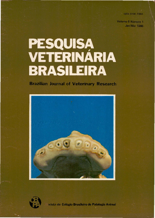
Dental lesions in cattle and sheep due to industrial pollution caused by coal combustion
- Abstracts: English Portuguese
Abstract in English:
Diverse dental lesions were verified on 11 farms located between 1.2 and 9.6 km from a coal-combustion thermoelectric plant in the municipality of Bagé, Rio Grande do Sul, Brazil. The incisor teeth of some animals appeared opaque with white spots, yellowishbrown discoloration and hypoplasia of the enamel. Gingival hyperplasia was also seen. These lesions were numerous on the farms close to the plant and less frequent on those more distant. The most important alteration was dental wear. Incisor teeth of cattle from farms near the plant were completely worn down by the time the animals were six to seven years of age; the degree of wear was related to the distance between the farms and the plant as a linear function [Y = 4.11 + (- 0.42x); r2 = 0.75 (P < O.OS)]. Histological lesions of permanent incisors were characterized by hyperplasia of the cernentum, proliferation of the dentin, and periodontal pocketing with alveolar bone resorption. Excessive and irregular wear was also observed in prernolar and molar teeth. Sheep presented lesions similar to those observed in cattle; ewes three to four years old showed completely worn down incisors. Fluoride levels in cattle on a farm located 2.45 km from the plant varied from 1091 to 5673 ppm (X̅ = 2539 ppm) in nine humerus bones and from 389 to 2931 ppm (X̅ = 1346 ppm) in 21 mandibles. These levels decreased as the distance from the plant increased; on a farm 7.5 km away, fluoride levels were 386 ppm in humerus bones and 265 ppm in mandibles. The results obtained confirm the diagnosis of fluoride poisoning, a condition that was previously described in the city of Rio Grande, Rio Grande do Sul, and attributed to pollution cause d by phosphate processing factories. In the Candiota region the enamel lesions were less pronounced and the fluoride levels lower than those observed in Rio Grande, however, the dental wear was much more accentuated in the animals from Candiota. Two factors might be responsible for this dental wear: the abrasive effect of particles eliminated into the air during coal combustion, the most important of which appears to be silica; and the decrease in the resistance of the enamel as a consequence of fluoride poisoning.
Abstract in Portuguese:
Diversas lesões dentárias foram constatadas em 11 propriedades localizadas entre 1,2 e 9,6 km de urna usina termoelétrica no município de Bagé, Rio Grande do Sul. Os dentes incisivos de alguns animais apresentavam-se opacos, com manchas brancas, pigmentação amarelo-marrom, hipoplasia do esmalte. Hiperplasia da gengiva também ·foi observada. Tais lesões eram consideráveis nos estabelecimentos mais próximos da usina e discretas nos demais. A alteração mais importante era o desgaste dentário. Os bovinos dos estabelecimentos mais próximos da usina apresentavam desgaste completo dos seus incisivos aos 6 ou 7 anos de idade; o grau de desgaste esteve relacionado à distância entre os estabelecimentos e a usina como uma função linear Y = 4,11 + (- 0,42. x); r2 = 0,75 (P < 0,05). As lesões histológicas dos incisivos permanentes foram caracterizadas por hiperplasia do cemento, proliferação de dentina reacional, e formação de bolsa peridentária com reabsorção do osso alveolar. Desgaste excessivo e irregular foi observado também em dentes premolares e molares. Os ovinos apresentaram lesões similares às dos bovinos; ovelhas de 3 à 4 anos de idade apresentavam desgaste total de seus incisivos. Os níveis de flúor em bovinos, em um estabelecimento situado a 2,45 km da usina variaram de 1091 a 5673 ppm (X̅ = 2539 ppm) em nove úmeros e de 389 a 2931 ppm (X̅= 1346 ppm) em 21 mandíbulas. Os níveis de flúor diminuíram a medida que aumentava a distância da usina; em um estabelecimento situado a 7 ,5 km os níveis foram de 386 ppm em úmero e 265 ppm em mandíbula. Os resultados obtidos confirmam o diagnóstico de intoxicação por flúor, doença que foi diagnosticada anteriormente no município de Rio Grande, RS, como conseqüência da poluição causada por fábricas de adubo. Na região de Candiota as lesões do esmalte são menos pronunciadas e os níveis de flúor mais baixos que os observados em Rio Grande; o desgaste dentário, no entanto, é muito mais acentuado nos animais de Candiota. Dois fatores seriam responsáveis pelo desgaste dentário: o efeito abrasivo dos particulados eliminados com o efluente da combustão de carvão, dos quais o mais importante pareceria ser o silício; a diminuição da resistência do esmalte como conseqüência da intoxicação por flúor.

